The Aesthetic Detective: Decoding Facial Bumps
The Aesthetic Detective: Decoding Facial Bumps
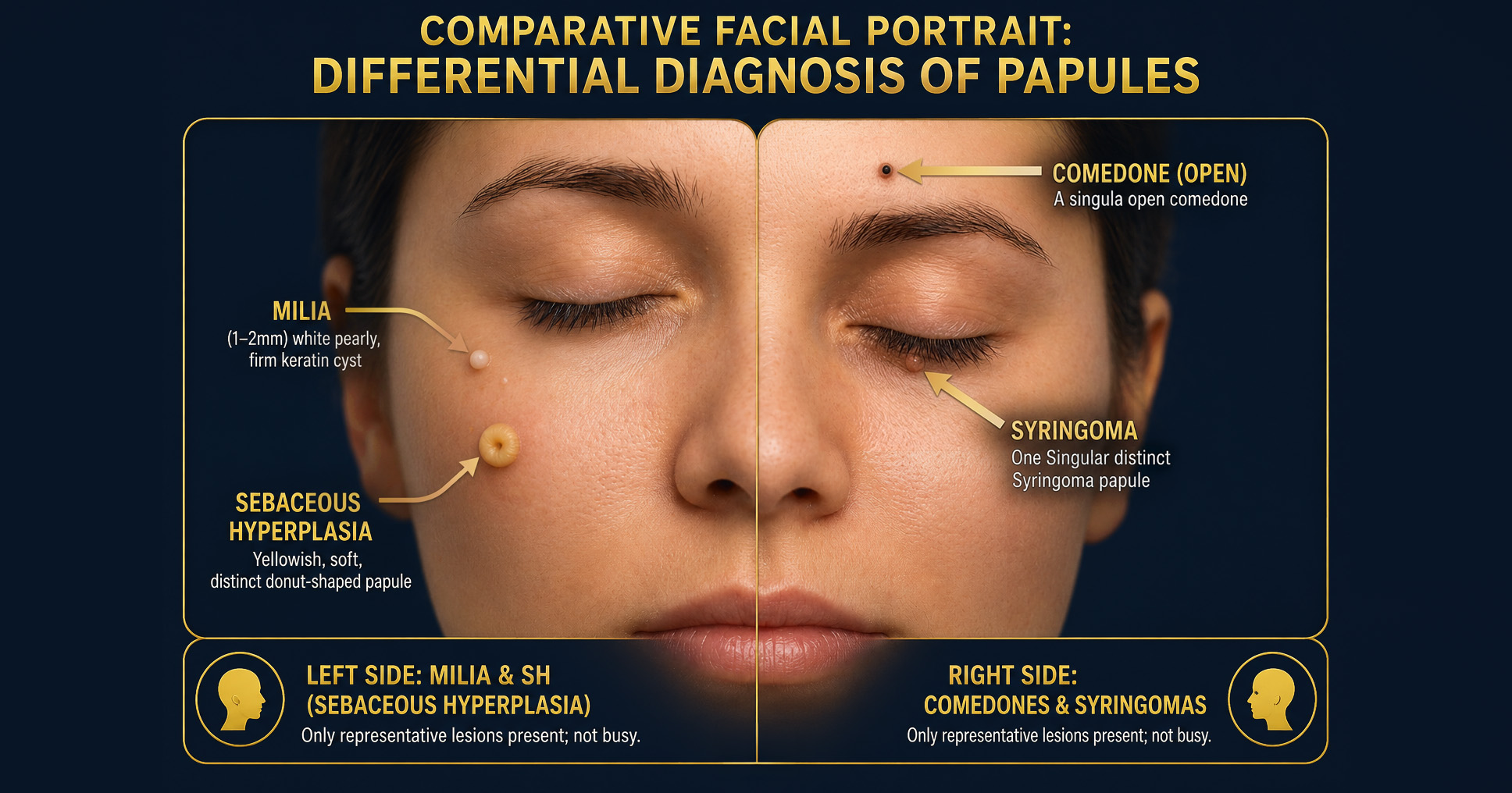
When shifting your clinical gaze from systemic emergencies to aesthetic dermatology, the focus moves from macro to micro. The face is a complex landscape, and to the untrained eye, every tiny bump looks like a pimple waiting to be popped. However, treating a benign facial lesion without understanding its underlying pathology leads to poor results, scarring, and frustrated patients. Here is the definitive guide to the differential diagnosis of common facial papules: Milia, Comedones, Sebaceous Hyperplasia, and the often confused Syringoma.
1. Milia: The "Skin Pearls"

Milia are tiny, superficial cysts located just under the epidermis. They are entirely walled off and contain no living tissue—just a hard, calcified-like ball of dead skin.
- Clinical Presentation: 1-2 mm, perfectly round, firm, bright white or pearly yellow domes. They have no visible pore opening.
- Causes: Occurs when keratin (skin protein) gets trapped beneath the surface. This can happen spontaneously (primary milia) due to sluggish cell turnover, or secondarily due to skin trauma, burns, blistering rashes, or the chronic use of heavy, occlusive creams.
- Prevention: Consistent chemical exfoliation (AHAs like Glycolic Acid) and topical retinoids to keep cellular turnover high and prevent dead skin from accumulating. Avoiding heavy, pore-clogging ointments, especially around the eyes.
- Treatment - Manual Extraction: Lancing the roof of the cyst with a sterile needle or scalpel (e.g., an 11-blade) and gently pressing out the keratin pearl with a comedone extractor.
- Treatment - Chemical Peels: Superficial peels (Glycolic or Salicylic acid) to thin the stratum corneum, making extractions easier or allowing them to resolve naturally.
2. Sebaceous Hyperplasia: The "Oil Donuts"

Sebaceous hyperplasia is a benign, chronic enlargement of the sebaceous (oil) glands. Unlike milia or comedones, this is living glandular tissue, not trapped debris.
- Clinical Presentation: 2-5 mm, soft, yellowish or flesh-colored papules. They classically feature a central dimple (umbilication) which represents the central follicular duct.
- Causes: Driven by genetics, chronic sun damage, and decreasing androgen levels associated with age. As cell turnover slows down, the sebaceous glands swell with trapped sebum and hypertrophy (overgrow), pushing up to the skin's surface.
- Prevention: Daily sunscreen use is critical to prevent the UV damage that exacerbates glandular changes. Long-term use of topical retinoids can help regulate gland function, though genetics play the biggest role.
- Treatment - Electrodessication: Using a fine-tipped hyfrecator to deliver a small electric current to the lesion, causing the overgrown tissue to contract and crust off. (Note: Do not extract. Squeezing will only yield a tiny bit of clear oil and leave the patient bruised. The gland itself must be destroyed or shrunk).
- Treatment - Laser Ablation: CO2 or Erbium lasers to vaporize the overgrown tissue.
- Treatment - Oral Medications: In severe, widespread cases, low-dose oral isotretinoin can radically shrink the glands.
3. Comedones: The "Clogged Pores"
Comedones are the primary lesions of acne vulgaris. They occur inside the hair follicle when sebum mixes with dead skin cells to create a plug.
- Clinical Presentation (Closed Comedones/Whiteheads): Small, flesh-colored or whitish bumps where the follicle is completely blocked, trapping the debris below the surface.
- Clinical Presentation (Open Comedones/Blackheads): The pore is stretched open. The dark color is not dirt; it is oxidized melanin and sebum exposed to the air.
- Causes: Hormonal fluctuations, excess oil production, hyperkeratinization (sticky skin cells), and comedogenic makeup or skincare products.
- Prevention: Daily cleansing with Salicylic Acid (BHA), non-comedogenic skincare routines, and topical retinoids (Tretinoin or Adapalene) to normalize follicular shedding.
- Treatment - Chemical Peels: Salicylic acid (20%-30%) is the gold standard here, as its lipophilic (oil-soluble) nature allows it to dive deep into the pore and dissolve the sebum plug.
- Treatment - Clinical Extractions: Safely pressing the debris out of the follicle using proper aesthetic technique.
4. The Tricky Look-Alike: Syringomas
When evaluating bumps around the eyes, Syringomas must be in your differential diagnosis. They are benign tumors of the eccrine (sweat) glands.
- Clinical Presentation: Clusters of small (1-3 mm), firm, flesh-colored or slightly yellowish papules, almost exclusively found on the lower eyelids and upper cheeks. They look remarkably similar to milia.
- Causes: Overactivity of the sweat glands, largely driven by genetics. They are more common in women and typically emerge in early adulthood.
- Prevention: Because they are structural glandular tumors driven by genetics, there is no effective topical prevention.
- Treatment Options: Like sebaceous hyperplasia, they cannot be extracted. They require tissue destruction via electrodessication, laser ablation, or deep TCA spot-treatment.